Peripheral Blood Cell Morphology and Interstitial Assessment —— Erythrocytes
Original Hou Bingbing Jing Wong Inspection Medical Network
Author: Hou Bingbing Jing Wong
SETTING: Shijiazhuang Traditional Chinese Medicine Hospital, Hebei Province
After the peripheral blood smear was stained with Wright’s stain, the normal red blood cells were pale pink (the center of the cell was a lightly stained area with a diameter of 1/3), and the size was uniform (6.7-7.7um). There were no other abnormal structures in the cells, and red blood cells were scattered at the junction of the body and tail of the blood smear. Various reasons may lead to changes in the morphology and structure of red blood cells, such as abnormalities in the size, shape, dyeing, structure and arrangement of red blood cells (note: human factors, such as production and dyeing, should be excluded first). The pictures in this article are mainly taken from Shanghai Clinical Laboratory Quality Control Center in previous years.
Macroerythrocytes
Macrocyte (diameter > 15um), macrocyte (diameter 10-15um). Erythrocyte volume changes are mostly seen in megaloblastic anemia caused by folic acid or vitamin deficiency, and also in hemolytic anemia or MDS and other diseases.
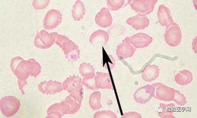
Target red blood cell
Red blood cells are larger in diameter, deeply stained in the center, lightly stained around and deeply stained at the edge of cells, such as shooting targets. It is more common in thalassemia. Such as timely drying and fixing when smearing can also cause similar changes.
Spiny red blood cell
The length, width and spacing of protrusions on the surface of red blood cells are different (pay attention to distinguish them from blunt sawtooth red blood cells). It is more common in β -lipoprotein deficiency (acanthocyte can reach 80%), and can be seen after splenectomy, alcoholic liver disease, uremia and so on.
Blunt sawtooth red blood cell
That is, shrunk red blood cells, the length, width and spacing of cell surface protrusions are the same (pay attention to distinguish them from blunt sawtooth red blood cells). More common in hypertonic environment or improper production.
cabot ring
Purple-red thin coil structure appearing in red blood cells, sometimes wound in a figure of eight. Its nature is not clear at present, and it is thought that it may be the residue of nuclear membrane or spindle or the product of lipoprotein denaturation in cytoplasm. Common in patients with megaloblastic anemia, hemolytic anemia, leukemia or lead poisoning.
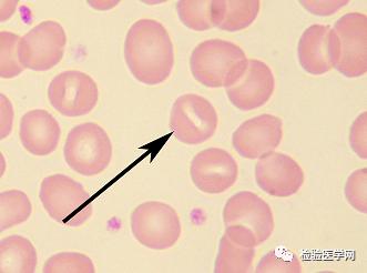
Mouth red blood cell
The pale area in the center of red blood cells is a narrow crack, the center of the crack is narrower than both ends, and the edge of the crack is clear, similar to a slightly open fish mouth. A small amount of oral red blood cells can also be seen in normal people’s peripheral blood, which is generally 3%-4%. If it exceeds 4%, it can be considered abnormal. When it increases, it is more common in hereditary oral polycythemia, which can be seen in DIC, alcoholism, hemolytic anemia and liver disease.
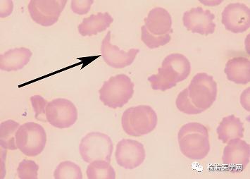
Helmet red blood cell
The shape is irregular, with 2 to 3 sharp corners in helmet shape, which is more common in disseminated intravascular coagulation, microangiopathic hemolytic anemia, cancer metastasis and so on.
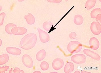
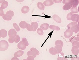
Teardrop red blood cell
Red blood cells are teardrop-shaped, pear-shaped, cudgel-shaped, etc., which are most common in myelofibrosis and hemolytic anemia.
schizocyte
The cells are irregular in shape, different in size, and have asymmetric pointed protrusions, which are red blood cell fragments or incomplete red blood cells. Usually irregular triangle, thorn or helmet shape. A small amount of erythrocytes can also be seen in normal people’s peripheral blood, which is generally less than 2%. If it exceeds 2%, it can be considered abnormal. It is a fragment of red blood cells caused by mechanical injury or endogenous abnormality, which is mainly found in DIC, microangiopathic hemolytic anemia, megaloblastic anemia, severe burn and so on.
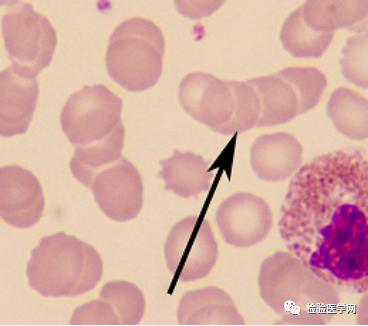
Money-shaped red blood cells
The arrangement of red blood cells, like copper coins strung together, indicates that the blood viscosity is high, and abnormal proteins appear in plasma, which reduces the negative charge of red blood cells, changes the plasma charge balance, and reduces the repulsive force between red blood cells, thus facilitating the formation of money-like aggregation and accelerating the erythrocyte sedimentation rate. More common in multiple myeloma, hyperglobulinemia, hyperfibrinemia and so on.
spherocyte
The lightly stained area in the center of red blood cells disappeared, and the whole cells were deeply and evenly colored. Under normal circumstances, a small amount of spherical red blood cells can also be seen in peripheral blood, generally less than 3%. The increase in the proportion of spherical red blood cells is more common in hereditary spherocytosis (often more than 25%, sometimes reaching 100%), autoimmune hemolytic anemia, abnormal hemoglobinopathy, liver cirrhosis, hypersplenism, chronic infectious diseases, globin body hemolysis, neonatal hemolysis and hemolytic diseases caused by erythrocyte enzyme deficiency.
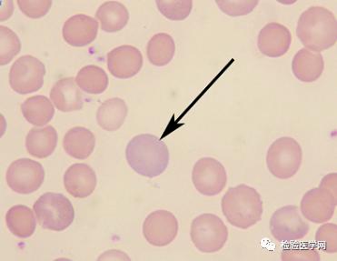
Polychromatic erythrocytes
The whole red blood cell or a part of its cytoplasm is grayish blue (due to the coexistence of intracellular RNA and hemoglobin), which is called polychromatic red blood cell. Under normal circumstances, a small amount of polychromatic erythrocytes can also be seen in peripheral blood, which is generally less than 1.5%. The increase of polychromatic erythrocyte ratio is more common in hemolytic anemia or acute hemorrhagic anemia, which indicates that bone marrow proliferative ability is active.
Basophilic stippling erythrocytes
Small blue particles with different sizes and scattered distribution can be seen in red blood cells, which is a relatively naive red blood cell. It may be due to the degeneration, aggregation and precipitation of basophils remaining in the cytoplasm after heavy metal damage to cells and the formation of particles. Under normal circumstances, basophilic spotted red blood cells (about 1/10000) are rarely seen in peripheral blood, but they are more common in heavy metal poisoning (such as lead, mercury, bismuth, etc.).
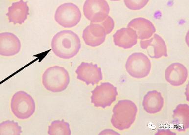
Howell-Jolly corpuscles
Red blood cells contain 1-2um purplish red round corpuscles with different sizes and numbers. This corpuscle may be the nuclear chromatin remaining during the division of young red blood cells. Common in hemolytic anemia, megaloblastic anemia, splenectomy, erythroleukemia or other proliferative anemia.
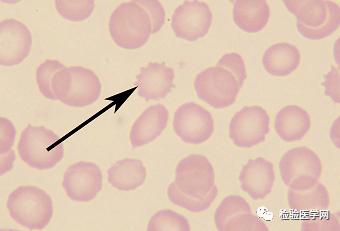
Oval red blood cell
Red blood cells are oval or rod-shaped, with a length of 3-4 times greater than the width, a maximum diameter of 12.5μm and a transverse diameter of 2.5 μ m.. This kind of red blood cell is placed in hypertonic, isotonic and hypotonic solution or normal human serum, and its oval shape remains unchanged, but neither young red blood cell nor reticulocyte is oval. Under normal circumstances, oval red blood cells are rarely seen in peripheral blood (about 1%, not more than 15%). In the blood smear of patients with hereditary ovalocytosis, the proportion of such red blood cells can reach 25% or even as high as 75%, and the proportion of oval red blood cells can also increase in thalassemia, megaloblastic anemia, myelosclerosis and iron deficiency anemia.
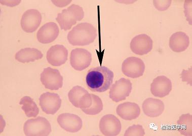
Nucleated red blood cell
Under normal circumstances, nucleated red blood cells can not be seen in adult peripheral blood, but a small number of nucleated red blood cells can be seen in neonatal peripheral blood within one week of birth. When nucleated red blood cells appear in adult peripheral blood, they can be seen in various proliferative anemia (hemolytic anemia, acute hemorrhagic anemia, megaloblastic anemia and severe hypopigmentation anemia can also appear), malignant tumor or metastasis of hematopoietic system, splenectomy and severe hypoxia.
Crescent red blood cell
Red blood cells are pale, incomplete and crescent-shaped (note that they are differentiated from sickle-shaped red blood cells). Mainly seen in some hemolytic anemia (such as PNH).
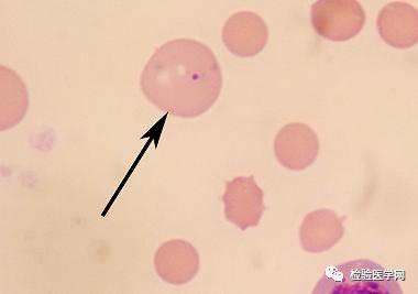
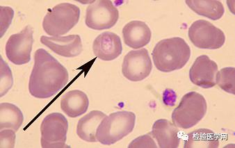
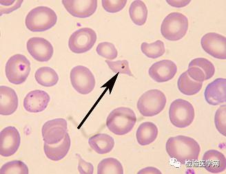
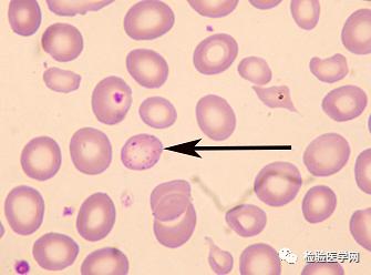
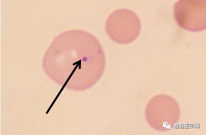
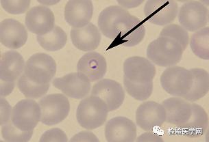
Red blood cells are infected with plasmodium (ring)
The nucleus of plasmodium ring is located on one side of the worm, which is shaped like a ring inlaid with precious stones. It is found in the peripheral blood of patients infected by plasmodium.
[References]
1. Wu Xiaoman. Basis of Clinical Laboratory [M]. 1st Edition. Wuhan: Huazhong University of Science and Technology Press, 2013.
Read the original text